Definition of Centrioles
- Near the nucleus, eukaryotic cells include two cylindrical, rod-shaped microtubular structures called centrioles.
- They are found in most algal cells (with the exception of red algae), moss cells, some fern cells, and most mammal cells, and lack a limiting membrane and DNA or RNA.
- Prokaryotes, red algae, yeast, cone-bearing and flowering plants (conifers and angiosperms), and some non-flagellated or non-ciliated protozoans are all devoid of them (such as amoebae).
- During mitosis or meiosis, centrioles form a microtubule spindle, the mitotic apparatus, and are sometimes positioned just under the plasma membrane to create and bear flagella or cilia in flagellated or ciliated cells.
- The basal body is the part of a centriole that has a flagellum or cilium.
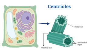
Figure: Centrioles in Plant Cell, Image Created with Bio Render
Centrioles structure
- Centrioles and basal bodies are cylindrical structures with a diameter of 0.15–0.25 um and a length of 0.3–0.7 um, though they can be as short as 0.16 um and as long as 8um.
- They can be seen under a light microscope, but only an electron microscope can disclose the complexities of the centriole structure.
- In the centrosome, a region near the nucleus, each cell has a pair of centrioles. Members of each pair of centrioles are at right angles to one another.
- They are sub-microscopic microtubular sub cylinders with a configuration of nine triplet fibrils with the ability to generate duplicates, astral poles, and basal bodies on their own, without the need for DNA or a membrane covering.
- A centriole has nine peripheral fibrils in its whorl. The core is devoid of fibrils. As a result, the configuration is known as 9 + 0. Fibrils run parallel to one other but at a 40-degree inclination. Three sub-fibers make up each fibril. As a result, it’s known as a triplet fibril.
- The three sub-fibers are actually microtubules that are connected by their edges and hence share the common walls of 2-3 proto-filaments.
- Each sub-fiber is 25 nm in diameter. The three sub-fibers of a triplet fibril are labelled C, A, and B from outside to inside. Due to the sharing of some microfilaments, sub-fiber A is complete with 13 proto-filaments, whereas and sub-fibers are incomplete.
- —A proteinaceous linkers join the adjacent triplet fibrils. The hub is a rod-shaped proteinaceous substance found in the core of centrioles. The hub is 2.5 nm in diameter. 9 proteinaceous strands extend from the hub to the periphery triplet fibrils. They’re known as spokes.
- Before joining with the A sub-fiber, each spoke has an X thickening. Nearby, there is another thickening called as Y. It is connected to both the X thickening and the C – A linkers via connectives.
- The centriole in T.S. has a cartwheel look due to the presence of radial spokes and peripheral fibrils.
Centrioles Functions
- The creation of the spindle apparatus, which acts during cell division, is aided by centrioles.
- Centrioles are missing in the mitotic process, causing divisional mistakes and delays.
- Each cilium or flagellum has a single centriole that serves as the anchor point or basal body.
- The production of cilia and flagella is also directed by basal bodies.
Click Here for Complete Biology Notes
Centrioles Citations
- Stephen R. Bolsover, Elizabeth A. Shephard, Hugh A. White, Jeremy S. Hyams (2011). Cell Biology: A short Course (3 ed.).Hoboken,NJ: John Wiley and Sons.
- Alberts, B. (2004). Essential cell biology. New York, NY: Garland Science Pub.
- Winey, M., & O’Toole, E. (2014). Centriole structure. Philosophical transactions of the Royal Society of London. Series B, Biological sciences, 369(1650), 20130457.
- http://www.biologydiscussion.com/cell/centrioles/centrioles-structure-and-functions-with-diagram/70541.
Related Posts
- Phylum Porifera: Classification, Characteristics, Examples
- Dissecting Microscope (Stereo Microscope) Definition, Principle, Uses, Parts
- Epithelial Tissue Vs Connective Tissue: Definition, 16+ Differences, Examples
- 29+ Differences Between Arteries and Veins
- 31+ Differences Between DNA and RNA (DNA vs RNA)
- Eukaryotic Cells: Definition, Parts, Structure, Examples
- Centrifugal Force: Definition, Principle, Formula, Examples
- Asexual Vs Sexual Reproduction: Overview, 18+ Differences, Examples
- Glandular Epithelium: Location, Structure, Functions, Examples
- 25+ Differences between Invertebrates and Vertebrates
- Lineweaver–Burk Plot
- Cilia and Flagella: Definition, Structure, Functions and Diagram
- P-value: Definition, Formula, Table and Calculation
- Nucleosome Model of Chromosome
- Northern Blot: Overview, Principle, Procedure and Results


YOURURL.com
Fox.wallet
mexican pharmacies that ship
canada pharmacy online
rhine inc viagra
free trial viagra sample
можно ли купить настоящий диплом можно ли купить настоящий диплом .