Definition of Bright field Microscope
The Compound Light Microscope is other name for the Bright field Microscope. It is an optical microscope which produces a dark image against a brilliant background by using light rays. It is the most common type of microscope used in Biology, Cellular Biology, as well as Microbiological Laboratory investigations.
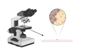
Bright field Microscope is used to examine fixed as well as live specimens which have been stained with basic stains to create contrast among the image as well as the backdrop. It is uniquely built with magnifying glasses referred to as lenses which alter the specimen to create an image visible via the eyepiece.
Bright field Microscope Principle
A specimen should pass through a consistent beam of illuminating light in order to be the focal point as well as create an image under the Bright field Microscope. The microscope will provide a contrasted image due to differential absorption as well as differential refraction.
The specimens are first stained to introduce colour for simple constricting characterisation. The coloured specimens will have a refractive index which distinguishes them from the neighboring environment, displaying a mix of absorption as well as refractive contrast.
The microscope’s operation is dependent on its ability to produce a high-resolution image from an adequately provided light source which is focused on the image, resulting in a high-quality image.
The specimen is seen on a microscopic slide while immersed in oil or covered with a coverslip.
Parts of Bright field Microscope
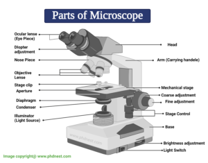
Figure: Parts of Brightfield Microscope (Compound Light Microscope)
Bright field Microscope Components
The bright field microscope is composed of several components, including:
- Eyepiece (Ocular lens) – The microscope contains two eyepiece lenses at the top which concentrate the image from the objective lenses. With your eyes, this is where you view the formed image.
- The objective lenses, which are composed of six or more glass lenses, produce a clean image of the specimen or object being focused.
- On the microscope’s arm are two focusing knobs, the fine adjustment knob as well as the coarse adjustment knob, which can be used to move the stage or the nosepiece to focus on the picture. Their job is to guarantee that the image produced is sharp as well as clear.
- The stage is located slightly below the objectives as well as is where the specimen is positioned, allowing movement of the specimen around for better viewing with the flexible knobs as well as where the light is concentrated.
- The condenser consists of the following components: It is positioned beneath the stage as well as focuses a light beam onto the specimen. To change the quality of light, it can be fixed or movable, although this is entirely dependent on the microscope.
- The arm is the solid metallic backbone of the microscope, used to transport as well as transfer it from one location to another. They also support the microscope base, which serves as the microscope’s stand. All of the microscopic components are held in place by the arm as well as the base.
- It has a light illuminator or a mirror at the base or on the nosepiece of the microscope.
- The nosepiece has two to five objective lenses with varying magnification power. To focus on the image, it can rotate to any position depending on the objective lens.
- The diameter of the light beam which passes through the condenser is controlled by an aperture diaphragm (contrast). When the condenser is almost closed, light passes through to the centre of the condenser, resulting in a high contrast image, whereas when the condenser is extensively open, the image is very bright with a low contrast.
Bright field Microscope Magnification
- The primary lenses used for focusing the picture on the condenser are the objective lenses. This results in an enlarged clear image, which is then magnified again by the eyepiece to form the primary image seen by the eyes.
- The objective lenses stay parfocal during imaging, which means that even if the objective lens changes, the image remains focussed. The image seen via the eyepiece is an enlarged clear image of the specimen, referred to as the virtual image.
- The image magnification is determined by comparing the magnification of the objective to the magnification of the eyepiece lens. Depending on the type of bright field microscope, the objectives have magnification powers ranging from 40x to 1000x, while the eyepiece lens has a basic magnification power of 10x.
As a result, to calculate:
Total magnification power = objective lens magnification x eyepiece magnification
- For example, if the objective has a magnification of 45x as well as the eyepiece has a magnification of 10x, the total magnification of the specimen is 450x.
- The magnification is standard, i.e. neither too high or too low, and so will range between 40X as well as 100oX depending on the magnification power of the lenses.
- The objective lens enlarges the image which can be seen, which is referred to as resolution. According to Prescott, resolution is the ability of a lens to differentiate or discern between small objects which are closely coupled together.
- While the eyepiece multiplies the image at the end of the viewing, its magnification range is lower than that of the objective lens at 8X-12X (10X standard) as well as 40X-100X, the magnification as well as resolution of the microscope are greatly dependent on the objective lens.
Bright field Microscope Applications
Brightfield Microscopes are utilised in a variety of fields, ranging from basic biology to comprehending cell structures in cell Biology, Microbiology, Bacteriology, as well as parasitic organism visualisation in Parasitology.
The majority of the specimens to be seen are stained with specific staining to allow for viewing. Negative staining as well as Gram staining are two of the staining techniques employed.
Some of its applications are as follows:
- Animal cells are visualised as well as studied using this technique.
- Plant cells are visualised as well as studied using this technique.
- It is used to investigate as well as visualise the morphologies of bacterial cells.
- Parasitic protozoans such as Paramecium are identified using this term.
Benefits of a Bright field Microscope
- It is easy to use, with only a few modifications required while viewing the image.
- We can use this to see both stained as well as unstained surfaces.
- The microscope’s optics have no effect on the colour of the specimen.
- The microscope can indeed be altered and altered for improved viewing, such as by inserting a camera to create a digital microscope or modifying the way of image lighting, such as by applying fluorochromes on the specimen and viewing in a dark environment to create a darkfield microscope.
Disadvantages of Bright field Microscope
- Because the aperture diaphragm might generate excessive contrast, which can affect the image’s outcome, the iris diaphragm is chosen.
- It is not suitable for viewing living specimens such as bacterial cells. Under the bright field microscope, only fixed specimens may be examined.
- The maximum magnification of a bright field microscope is 100x; however, modifications can boost the magnification to 1000x, which is the ideal magnification for bacterial cells.
- Because of the poor contrast, many specimens should be dyed in order to be seen.
- Visual distortion may result from the usage of oil immersion.
- Using a coverslip can result in specimen damage.
- Staining can inject superfluous undesirable features into the specimen or pollute it.
- It is time-consuming to dye the specimen before seeing it using a bright field microscope.
- For enlargement, the microscope needs a strong source of light, and the source of light can sometimes generate a great deal of heat, that might harm or kill the specimen.
Brightfield Microscope Citations
- Image created using Biorender.com
- https://www.microscopemaster.com/brightfield-microscopy.html
- https://www.thomassci.com/scientific-supplies/Brightfield-Microscope
- https://www2.hawaii.edu/~johnb/micro/m140/syllabus/week/handouts/m140.2.4.html
- https://www.prior.com/wp-content/uploads/2017/07/PriorLuxPOL-Manual.pdf
- https://www.coursehero.com/file/p4i3koh/Ocular-The-total-magnification-of-an-image-is-determined-by-and-The-magnifica/
- https://www.answers.com/Q/What_is_the_total_magnification_of_a_microscope
- http://www.tpub.com/corpsman/230.htm
- https://www.microscope.com/compound-microscope-parts/
- https://www.chegg.com/homework-help/questions-and-answers/list-three-types-specimens-dissecting-microscope-might-used-view-microscope-structures-use-q15195231
- https://quizlet.com/85147377/microscope-flash-cards/
Related Posts
- Phylum Porifera: Classification, Characteristics, Examples
- Dissecting Microscope (Stereo Microscope) Definition, Principle, Uses, Parts
- Epithelial Tissue Vs Connective Tissue: Definition, 16+ Differences, Examples
- 29+ Differences Between Arteries and Veins
- 31+ Differences Between DNA and RNA (DNA vs RNA)
- Eukaryotic Cells: Definition, Parts, Structure, Examples
- Centrifugal Force: Definition, Principle, Formula, Examples
- Asexual Vs Sexual Reproduction: Overview, 18+ Differences, Examples
- Glandular Epithelium: Location, Structure, Functions, Examples
- 25+ Differences between Invertebrates and Vertebrates
- Lineweaver–Burk Plot
- Cilia and Flagella: Definition, Structure, Functions and Diagram
- P-value: Definition, Formula, Table and Calculation
- Nucleosome Model of Chromosome
- Northern Blot: Overview, Principle, Procedure and Results

