Anatomy of Flowering Plants
Anatomy of Flowering Plants: Plant anatomy or phytotomy is the general term for the study of the internal structure of plants. Originally it included plant morphology, the description of the physical form and external structure of plants, but since the mid-20th century plant anatomy has been considered a separate field referring only to internal plant structure. Plant anatomy is now frequently investigated at the cellular level, and often involves the sectioning of tissues and microscopy.
Anatomy –Study of internal structure.
Flowers are not just pretty things to look at. Other than their beauty and fragrance they fulfil many more important functions. To start with, they are the reproductive organ of plants. They consist of many structures that help the plant survive, grow and reproduce. Let us take a look at the anatomy of flowering plants.
Cells with the same structure and functionality constitute a tissue
1 . Meristematic Tissue-
Growth is mainly done by regions of active cell division- meristems, which occur at the tips of roots and shoots.
- Apical Meristem – occur at the tip of root & shoot and produce primary tissue.
Axillary bud – During the formation of leaves and elongation of the Stem, some cells left behind from shoot apical Meristem, present in axils of leaves and can form branch/flower.
- Intercalary Meristem – occur between mature tissues (grasses & regenerate parts removed by grazing herbivores)
These both constitute primary Meristem.
- Secondary Meristem – Occur in mature region of roots & shoots, produce woody axis, cylindrical. E.g.- Fascicular vascular cambium, interfascicular cambium, cork cambium).
2. Permanent tissue –
When nearly formed, cells become structurally and functionally specialized and lose the ability to divide.
Simple tissue – All cells are similar in structure and function.
- Parenchyma- Major component, isodiametric, may be oval or spherical or round, walls are thin & cellulosic, closely packed with fewer spaces, storage secretion, photosynthesis.
- Collenchyma – Below epidermis, either homogenous layer/patches, thickened at corners due to cellulose, hemicellulose, pectin, may be oval/spherical/polygonal, assimilate food when containing chloroplast, spaces (-), mechanical support.
- Sclerenchyma – Long, narrow cells with thick, lignified cell walls, usually dead (low protoplast), found in fruit walls of nuts, the pulp of guava.
Fibres – thick-walled elongated, pointed
Sclereids – Highly thickened, and spherical, narrow lumen.
3. Complex tissue – Different types of cells.
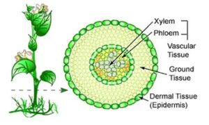
- Xylem – Water & mineral conduction from root to stem/leaves.
TRACHEIDS – elongated tube-like cells, thick lignified walls, tapering ends, dead, (+) in Gymnosperm for water conduction.
VESSELS – Cylindrical tube with many cells (vessel members), large central cavity, dead, interconnected through perforations, (-) in Gymnosperm.
FIBRES – Highly thickened walls, either septate or aseptate
PYRENCHYMA – Living, thin-walled with cellulose, store food in the form of starch or tannies, radial conduction of water takes place by ray parenchymatous cells.
Protoxylem – First formed primary xylem elements.
Metaxylem – Later formed.
Endarch – Protoxylem towards Centre, metaxylem towards the periphery. E.g., Stem
Exarch – Protoxylem towards the periphery, metaxylem towards Centre. E.g., Root
- Phloem – Transport food Material from leaves to other plants.
Sieve tube element – Long, tube-like, longitudinally arranged, end walls are perforated in a sieve-like manner to form sieve plates; the function is controlled by the nucleus of companion cells as they lack it, absence in gymnosperms.
Companion cells – Parenchymatous cells maintain pressure gradient in sieve tubes, absence in gymnosperms.
Parenchyma – Elongated, tapering cylindrical cells, dense cytoplasm, a cellulosic wall with pits for plasmodesmata connections, stores resins, latex, mucilage, (-) in monocots.
Fibres – Schlerenchymatous cells, absence in primary phloem, elongated, unbranched, pointed, thick wall, commercially – jute, flax, hemp.
Protophloem – First formed with narrow sieve tubes
Metaphloem – Later formed with bigger sieve tubes.
TISSUE SYSTEM
Epidermal tissue system –
Forms outermost covering of whole plant body, comprises- epidermal cells, stomata, appendages.
- Epidermis – Outermost layer, elongated, compactly arranged, single-layered, parenchymatous, large vacuole, covered with thick waxy layer- cuticle (prevent water loss), (-) in roots(cuticle).
- Stomata – Regulate transpiration & gaseous exchange, possess two bean-shaped cells – guard cells, dumbbell-shaped – grasses, an outer wall of guard cells is thin, inner- thick, possess chloroplast. Subsidiary cells – few cells near guard cells become specialized in shape and size: Stomatal apparatus- aperture, guard cell, subsidiary cells.
- Epidermal appendages – Cells of epidermis bear no. of hairs.
Root – root hairs (unicellular, absorb water & mineral)
Stem – Trichomes (multicellular, branched/unbranched, soft/stiff, prevent water loss).
Ground tissue System –
All tissue except epidermis & vascular bundle.
- Parenchymatous – in cortex, pericycle, pith & medullary rays.
- Collenchyma & sclerenchyma – In leaves, they consist of thin-walled chloroplast containing cell – mesophyll.
Click Here for Complete Biology Notes
Vascular tissue system –
- Radial – When xylem & phloem are arranged in an alternate manner along different radii. E.g., Roots
- Conjoint – Xylem & phloem are along the same radius of the vascular bundle. E.g., Stem and leaves.
The closed – Vascular bundle have no cambium, don’t show second growth. E.g., Monocots.
Open – Have cambium, show secondary growth. E.G., Dicots
Anatomy of dicot and monocot plants –
Dicot roots –
- Epiblema – Outermost layer, form hairs.
- Cortex – Thin-walled parenchyma cells, spaces (+), innermost layer – endodermis, single layer of barrel-shaped cells, (-) spaces, have a layer of the suberin (water impermeable) in the form of Casparian strips.
- Pericycle – few layers of thick-walled parenchymatous cells.
- Pith – small.
- Conjunctive tissue – between xylem & phloem (2-4)
- Stele – All tissue on the inner side of endodermis (pericycle, V.B., Pith)
Monocot roots –
- Similar to dicot root.
- It has more xylem bundles (more than 6)
- Pith is large & well developed
Dicot Stem –
- Epidermis – Outermost protective layer, covered with cuticle, trichomes.
- Cortex – Between epidermis & pericycle
Hypodermis – Collenchymatous cells, mechanical strength.
Cortical – Rounded thin-walled parenchymatous, (+) spaces
Endodermis – Innermost layer, rich in the starch grain – starch sheath
- Pericycle – inner side of endodermis, patches of sclerenchyma
- Medullary rays – Between vascular bundle, radially placed parenchymatous cells.
- Vascular bundles – In a ring, conjoint open, Endarch Protoxylem
- Pith – Rounded, parenchymatous cells with spaces in the Centre.
Monocot Stem –
- Similar to dicot stem.
- Hypodermis is sclerenchymatous
- Scattered vascular bundle, conjoint closed
- Phloem parenchyma (-)
Dorsiventral leaf –
Epidermis, mesophyll, vascular system.
- Epidermis – Both upper (adaxial) & lower (abaxial) surface with cuticle, abaxial has Ore stomata.
- Mesophyll – Tissue between upper & lower epidermis, parenchymatous.
Palisade parenchyma – adaxial, elongated, vertical cells, parallel.
Spongy parenchyma – Oval/round, loosely arranged.
- Vascular system – Vascular bundles in veins & mid -rib, surrounded by thick-walled bundle sheath cells.
Isobilateral (Monocot) leaf –
- Stomata are present on both surfaces.
- The mesophyll is not differentiated into palisade & spongy parenchyma.
- Bulliform cells – In grasses, adaxial cells modify into large, empty, colourless cells. When they have the water, they expose the leaf; when not, they curl the leaf to minimize water loss.
Secondary growth
Lateral meristem – vascular & cork cambium
Vascular cambium –
The meristematic layer is responsible for cutting off vascular tissue.
- Formation of the cambial ring –
Intrafascicular – cambium between primary xylem & phloem.
Interfascicular – Cells of medullary rays adjoining them become a meristematic, continuous ring of cambium.
- The activity of cambial ring –
The cambial ring becomes active & cut off new cells, both in inner & outer.
Secondary xylem – Cell cut off towards pith (matured)
Secondary phloem – Cells cut off towards periphery (matured).
Cambium is more active on the inner side so, Secondary xylem > Secondary phloem.
Secondary medullary rays – Cambium forms a narrow band of parenchyma passing through the secondary xylem & phloem in the radial direction.
- Springwood & Autumn wood –
Springwood – In spring, cambium is more active and produce more xylem having wider vessels, also called earlywood, low density, lighter colour.
Autumn wood – In winter, it is less active and produces less xylem with the narrow vessel, also called latewood, high density, dark colour.
Annual ring – The two kinds of wood appear as alternate concentric rings, gives an estimate of the age of the tree.
- Heartwood & Sapwood –
Heartwood – Region of Secondary xylem of dead elements with the lignified wall, doesn’t conduct water.
Sapwood – Peripheral region of Secondary xylem, lighter in colour, conducts water and mineral.
The secondary xylem is dark due to tannins, resins, oil, gums which make it hard, durable, resistant to microbes.
Cork Cambium-
- Cork cambium/Phellgen – As Stem continues to increase in girth due to vascular cambium, outer cortical and epidermis layer get broken, so new meristematic tissue is formed, couple of layers thick, narrow, thin-walled rectangular cell, it cuts off cells on both sides.
- Cork/phellem – outer cells differentiate into phellem.
- Secondary cortex/ Phelloderm – Inner cells differentiate into phelloderm.
- PERIDERM – Phellogen + Phellem + Phelloderm
- Bark – When periderm die and slough off, tissues exterior to the vascular cambium.
Early/soft bark – Formed early in the season
Late/hard – End of season.
- Lenticels – Phellogen cuts off closely arranged parenchymatous cells on the outer side instead of cork cells. This Rupture is forming a lens-shaped opening that permits the exchange of gases that occurs in woody trees.
Secondary growth in roots –
Vascular cambium originates from the tissue below phloem bundles, a portion of pericycle tissue above protoxylem forming a complete continuous wavy ring that becomes circular.
Related Posts
- Phylum Porifera: Classification, Characteristics, Examples
- Dissecting Microscope (Stereo Microscope) Definition, Principle, Uses, Parts
- Epithelial Tissue Vs Connective Tissue: Definition, 16+ Differences, Examples
- 29+ Differences Between Arteries and Veins
- 31+ Differences Between DNA and RNA (DNA vs RNA)
- Eukaryotic Cells: Definition, Parts, Structure, Examples
- Centrifugal Force: Definition, Principle, Formula, Examples
- Asexual Vs Sexual Reproduction: Overview, 18+ Differences, Examples
- Glandular Epithelium: Location, Structure, Functions, Examples
- 25+ Differences between Invertebrates and Vertebrates
- Lineweaver–Burk Plot
- Cilia and Flagella: Definition, Structure, Functions and Diagram
- P-value: Definition, Formula, Table and Calculation
- Nucleosome Model of Chromosome
- Northern Blot: Overview, Principle, Procedure and Results

