Definition of Endocytosis
- Endocytosis is a cellular mechanism that allows a cell to internalise substances from its surroundings, such as proteins, electrolytes, fluids, microorganisms, and some macromolecules. These compounds are broken down into smaller elements by a variety of operations, either for use by the cell or for waste.
- The immune system’s white blood cells are perhaps the most prevalent cells which make use of endocytosis to remove microbial infections from the body. They trap, break down, and wipe out viruses so that they can be eliminated from the body.
- The discovery of cellular elements like lysosomes, peroxisomes, endosomes, as well as exocytosis cellular mechanisms, together with endocytosis, was initially explained by Christian de Devu, a Belgian cytologist and biochemist who received numerous Nobel prizes for his role in the discovery of cellular elements like lysosomes, peroxisomes, endosomes, as well as exocytosis cellular mechanisms, including the endocytosis mechanism.
Endocytosis Process
- Invagination occurs when the cell membrane folds, generating a space filled with extracellular fluid, dissolved chemicals, food particles, foreign objects, pathogens, and/or other things.
- Following invagination, the cell membrane folds back on itself to form an evenly enclosed membrane surrounding the trapped molecules, which is referred to as a vesicle. Into the cell cytoplasm, some cells generate extension channels.
- The newly produced vesicle separates from the cell membrane and is processed by the cell.
Endocytosis Mechanisms Types
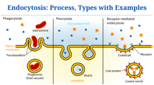
Endocytosis can be classified into three types:
- Phagocytosis
- Pinocytosis
- Endocytosis Induced by Receptors (Clathrin-Mediated Endocytosis)
-
Phagocytosis
- Phagosome formation occurs when a cell’s cell membrane spreads in the direction of a particle, surrounding it in addition to confining it in a folded membrane to form a phagosome. The substance swallowed by the phagosome is then fined by cellular enzymes. White blood cells (macrophages, monocytes, neutrophils, Eosinophils, and dendritic cells) within multicellular creatures use phagocytosis to eliminate pathogens. Phagocytosis is used by a few protozoans, like Entamoeba spp., to absorb nutrition.
- William Osler, a Canadian physician, identified the mechanism of phagocytosis (1876).
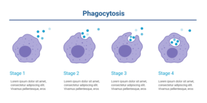
Phagocytosis is divided into five stages:
- Phagocytic cells recognise and move towards the antigen or chemical of interest.
- After then, the phagocyte binds to the antigen or target molecule. Phagocytes have the ability to surround the pathogen’s target particle by extending their membranes (pseudopodia). While encapsulating the particles, the pseudopodia are pointing in the same direction. The particle is after that contained by vesicle created by fused extended pseudopodia.
- A phagosome is a vesicle with particles encapsulated within it. This is the vesicle that perhaps the phagocyte consumes.
- The phagosome joins phagocyte’s lysosomes to produce a phagolysosome. The contents of the vesicle are degraded or digested by digestive enzymes in the lysosomes.
- Exocytosis is the process of removing degraded particles from phagocytic cells.
Figure: Stages of Phagocytosis image created with BioRender
Phagocytosis Example
- The mechanism of phagocytosis is well illustrated by immune cells like macrophages, dendritic cells, and neutrophils. Macrophages are immune system’s biggest phagocytic cells. They work by recognising, adhering to, consuming, digesting, and exocytosing digested particles out of the cytoplasm. Bacteria, fungus, dust particles, dead cells, and other antigens are among them. Macrophages are the immune system’s main phagocytic cells. A pseudopodial membrane is present. When they detect an antigen, they migrate toward it, extending their pseudopodia toward it, and engulfing it. When a cell is engulfed by an antigen, it produces a phagosome, which is a cell vesicle that ingests the antigen. The vesicle collides with lysosomes within the macrophage, generating a phagolysosome, that is broken down by lysosomic enzymes, subdividing the particle, that will be exocytosed out of the cell.
- The chemical composition of the particle has a significant impact on its ability to adhere to the phagocyte. Some bacterial antigens bind directly to the phagocyte, others such as complements of antibodies, need the creation of a film on the bacterial surface by a protein component from the blood called an opsonin, a process known as opsonization. The phagocytes must initially attach to the opsonin before phagocytosis may occur.
- Even with an opsonin, a few bacteria having enclosed cell walls are hard to digest. As a result, after the body reacts to their existence, they must be attached to certain antibodies. The phagocytes can then act on the antibody-bound encapsulated bacteria.
-
Pinocytosis
- Cell drinking is also known as fluid endocytosis. This type of endocytosis is an endocytosis in which tiny particles from external fluids infiltrate the cell membrane, resulting in the formation of a tiny vesicle containing suspended tiny molecules or particles inside the cell. The pinocytic vesicle joins the cell endosome for particle digestion.
- The main disparity among pinocytosis and phagocytosis is that in pinocytosis, ingested particles are restricted to the extracellular fluids of the cell. By invaginating the vesicle having the fluid particles, the cell membrane was able to transfer them into the cell lysosomes. When the vesicle and the lysosomes combine, the lysosomes release digesting enzymes. The enzymes break down the vesicle, discharging its contents into the cytoplasm for the use of the cell.
- Vesicles don’t always engage with lysosomes; as an alternative, they travel across the cell, merging with the cell membrane, causing membrane proteins and lipids to be recycled.
- There are two processes that cause pinocytosis:
(a) Micropinocytosis – It is the creation of tiny vesicles with a diameter of around 0.1 um; it occurs in body cells and results in caveolae, which are small growing vesicles on the cell membrane. They ae present inside the endothelium of blood vessels.
(b) Macropinocytosis – The cell membrane ruffles (villi), that are projections which expand to the extracellular fluids and include the capability to fold back on their own, generate the enormous vesicles. White blood cells contain them. While folding, they shove extracellular fluid into the cell, making a vesicle which draws into the cell.
Pinocytosis example
Pinocytosis can be shown in the following example
Nutrient absorption or ingestion inside the small intestines.
- Receptor Mediated Endocytosis (Clathrin Mediated Endocytosis) This kind of endocytosis is also referred to as clathrin-mediated endocytosis. It engages the internalisation in addition to recycling of receptors involved in signal transduction (G-protein and tyrosine kinase receptors), food intake, and synaptic vesicle reformation.
- The process of Receptor Mediated Endocytosis (Clathrin Mediated Endocytosis) is commenced by the gathering of phosphatidylinositol-4,5-bisphosphate (PIP2) inside the cell membrane. PIP2 is produced by catalysis of phosphoinositide in the plasma membrane. When phosphoinositide is converted to phosphoinositide by lipid kinase and phosphatases are hydrolyzed, PIP2 is produced.
- Clathrin-coated vesicles (CCV) should invaginate as well as develop in order to produce clathrin-coated pits. Clathrin-coated vesicles connect to the cell membrane, attracting a range of proteins involved in vesicle maturation, such as Actin-binding proteins and Adapter proteins (AP).
- With the support of dynamin protein, the CCV invaginates into the cell membrane, develops, and splits from the cell membrane, making a clathrin-coated pit.
- What causes the formation of Clathrin-coated pits? Because clathrin-coated vesicles are prevalent in nearly all the cells, when a signal is detected, these vesicles employ the adaptor proteins in the plasma membrane, which cluster on the lipid layer of the plasma membrane.
- Adaptor proteins include clathrin from clathrin-coated vesicles, as well as Actin-binding proteins, into the cell membrane lipids. Lipids layer’s negative charge attracts the clathrin’s positive charge, resulting in a concave shape that rises over the membrane, making pits all over the plasma membrane.
- On the pits, the clathrin behaves like a sensor for endocytosis-inducing signals, whereas the Clathrin Coated Vesicles’ vesicle is recycled back to the cell membrane.
- The cycle among clathrin-coated pits and clathrin-coated vesicles generation will continue so long as signalling receptors and ligands which trigger them exist.
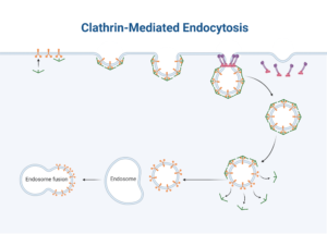
- The steps in the receptor-mediated endocytosis process are as follows:
- The particles (ligands) which must be produced are linked to receptors on the cell membrane, which form coated pits when they cluster together. The pits then invaginate, generating a vesicle that, with the assistance of the dynamin proteins, pinches off within the cell membrane. The vesicles after that lose clathrin and adaptor proteins.
- When an uncoated vesicle joins an early endosome, the late endosome, also known as the sorting vesicle, is created. Within the vesicle, the late endosome removes the receptors from the ligands and recycles them into the cell membrane.
- When the particles are discharged, they come into contact with lysosomes, which have digestive enzymes which hydrolyze the content of vesicle. Following that, the digested particles are discharged into the cell for usage.
- Clathrin-coated endocytosis (receptor-mediated endocytosis) is the best way to get macromolecules into the cell.
Image created with BioRender
Exercising Clathrin-Mediated Endocytosis
- Two well-known examples of Clathrin-Mediated Endocytosis are:
- iron-bound transferrin recycling
- Receptor-mediated endocytosis is the uptake of cholesterol bound to low-density lipoprotein (LDL), a mix of phospholipid, protein, and cholesterol.
Click Here for Complete Biology Notes
Endocytosis Citations
- https://www.thoughtco.com/what-is-endocytosis-4163670
- https://bio.libretexts.org/Bookshelves/Cell_and_Molecular_Biology/Book%3A_Basic_Cell_and_Molecular_Biology_(Bergtrom)/17%3A_Membrane_Function/17.4%3A_Endocytosis_and_Exocytosis
- https://www.pathwayz.org/Tree/Plain/ENDOCYTOSIS+%26+EXOCYTOSIS
- https://www.britannica.com/science/pinocytosis
- MBINFO Defining Mechabiology:www.mechabio.info
- https://www.biologyonline.com/dictionary/opsonization
- https://www.britannica.com/science/phagocytosis
- https://www.britannica.com/science/pinocytosis
Related Posts
- Phylum Porifera: Classification, Characteristics, Examples
- Dissecting Microscope (Stereo Microscope) Definition, Principle, Uses, Parts
- Epithelial Tissue Vs Connective Tissue: Definition, 16+ Differences, Examples
- 29+ Differences Between Arteries and Veins
- 31+ Differences Between DNA and RNA (DNA vs RNA)
- Eukaryotic Cells: Definition, Parts, Structure, Examples
- Centrifugal Force: Definition, Principle, Formula, Examples
- Asexual Vs Sexual Reproduction: Overview, 18+ Differences, Examples
- Glandular Epithelium: Location, Structure, Functions, Examples
- 25+ Differences between Invertebrates and Vertebrates
- Lineweaver–Burk Plot
- Cilia and Flagella: Definition, Structure, Functions and Diagram
- P-value: Definition, Formula, Table and Calculation
- Nucleosome Model of Chromosome
- Northern Blot: Overview, Principle, Procedure and Results

