Eosin Methylene Blue (EMB) Agar
Eosin Methylene Blue (EMB) agar is a microbiological selective stain that limits Gram-positive bacteria’s growth while simultaneously serving as a colour indication for identifying organisms which help ferment lactose (e.g., E. coli) even those who don’t (e.g., E. coli) (e.g., Salmonella, Shigella).
- EMB agar was invented by Holt-Harris and Teague, and was later customised by Levine.
- With Levine’s peptic digest of animal tissue with phosphate and Holt-Harris and Teague’s two carbs, it’s a hybrid of the Levine, Holt-Harris, and Teague formulations.
- This medium is essential in clinical laboratories for quickly differentiating gram-negative dangerous bacteria.
Compositions of EMB Agar
| Ingredients | Gms/litre |
| Agar | 13.500 |
| Peptic digest of animal tissue | 10.000 |
| Lactose | 5.000 |
| Sucrose | 5.000 |
| Dipotassium phosphate | 2.000 |
| Methylene blue | 0.065 |
| Eosin – Y | 0.400 |
Final pH (at 25°C): 7.2±0.2
Top Online PhD Programs : Pros and Cons, Universities, Courses, Career.
EMB Agar Principle
- Existence of a 6:1 integration of the two dyes eosin as well as methylene blue distinguishes EMB agar.
- Lactose fermentation by Gram-negative bacteria releases acid, which lowers the pH level. Because the acid reacts with the colours, it increases colony dye consumption, resulting in dark purple colonies.
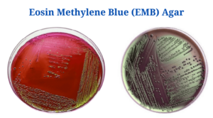
- Lactose-fermenting bacteria can also create flat, black colonies with a metallic green luster.
- A number of lactose fermenters create only larger, mucoid colonies with purple centres.
- The large percentage of E. coli colonies on EMB agar have quite a striking green sheen; lactose non-fermenters, on the other hand, can increase the pH through protein deamination, culminating in a quick drop in the pH of the EMB agar, that is a crucial factor in the production of the green metallic sheen shown in E. coli, and also quick lactose fermentation and the development of strong acids. This prevents the colour from being consumed. Colonies will have no colour.
- Lactose non-fermenters are light lavender or white in colour.
- The peptic digestion of animal tissue provides carbon, nitrogen, and other critical growth components.
- Lactose and sucrose are both fermentable carbohydrates that provide energy.
- Eosin-Y and methylene blue are examples of differential indicators. Phosphate is a medium-sized buffer.
Preparation and Method of Use of EMB Agar
- Dissolve 35.96 grammes in 1000 mL distilled water.
- Blend till the suspension is smooth as well as consistent. Bring the medium to a boil to completely dissolve it.
- Autoclave for 15 minutes at 15 lbs pressure (121°C) to sterilise. AT ALL COSTS, PREVENT OVERHEATING.
- Cool to 45-50°C and stir the medium to oxidise the methylene blue (restore its blue hue) while retaining the flocculent precipitate.
- Pour the mixture into sterile Petri plates.
- Bring the plates to room temperature and then use.
- Before inoculating, make sure the agar surface is fully dry.
- As quickly as possible following collection, innoculate and stripe the specimen.
- Roll the object which has to be cultivated over a tiny space of the agar surface and streak it with a sterile loop to isolate it.
- Incubate aerobically for 18-24 hours at 35-37°C while shielding plates from light.
- Examine the colonies on the plates for morphology. If somehow the findings are negative after 24 hours, incubate for yet another 24 hours.
Result Interpretation on EMB Agar
| Organisms | Growth |
|
Enterobacter aerogenes |
Fine development; pink, with no shine |
| Escherichia coli | Blue-black bulls eye; might contain green metallic shine |
| Pseudomonas aeruginosa | Without any colour |
| Klebsiella pneumoniae | Pink, mucoid colonies |
| Proteus mirabilis | Abundant development; without any colour colonies |
| Salmonella Typhimurium | Abundant development; without any colour colonies |
Click Here for Complete Biology Notes
Uses of EMB Agar
- For the isolation and differentiation of gram-negative enteric bacteria from clinical and nonclinical specimens, EMB Agar (Eosin Methylene Blue Agar) is indicated.
- It allows us to distinguish between gram-positive and gram-negative microorganisms.
- It helps in the isolation and separation of gram-positive and gram–negative bacteria.
- This is used to evaluate the water quality, particularly to see if it’s polluted with hazardous germs.
- It distinguishes bacteria from the colon, typhoid, and dysentery groups.
- Escherichia coli, various nonpathogenic lactose-fermenting enteric gram-negative rods, and the Salmonella and Shigella genera can all be distinguished using EMB media.
- This also aids in the differentiation of lactose-fermenting versus non-lactose-fermenting enteric bacteria.
Limitations of EMB Agar
- In addition to EMB Agar, a non-selective medium must be utilised.
- For comprehensive identification, biochemical, immunological, molecular, or mass spectrometry testing on colonies from pure culture is indicated.
- Some Salmonella and Shigella strains may not be appropriate for EMB Agar.
- Gram-positive bacteria such as enterococci, staphylococci, and yeast can grow on the medium and form pinpoint colonies.
- In this environment, non-pathogenic, non-lactose-fermenting microbes will grow. To identify these species from those that are harmful, additional biochemical testing are required.
- For efficient isolation of mixed flora samples, serial inoculation may be required.
- Because certain strains of E. coli do not exhibit an unique green metallic sheen, the green metallic sheen is not diagnostic for E. coli.
Eosin Methylene Blue Citations
- https://microbeonline.com/eosin-methylene-blue-emb-agar-composition-uses-colony-characteristics/
- http://himedialabs.com/TD/M317.pdf
- https://microbeonline.com/eosin-methylene-blue-emb-agar-composition-uses-colony-characteristics/
- https://catalog.hardydiagnostics.com/cp_prod/Content/hugo/EMBAgar.htm
- https://bio.libretexts.org/Ancillary_Materials/Experiments/Microbiology_Labs_I/23%3A_Eosin_Methylene_Blue_Agar_(EMB)
Related Posts
- Phylum Porifera: Classification, Characteristics, Examples
- Dissecting Microscope (Stereo Microscope) Definition, Principle, Uses, Parts
- Epithelial Tissue Vs Connective Tissue: Definition, 16+ Differences, Examples
- 29+ Differences Between Arteries and Veins
- 31+ Differences Between DNA and RNA (DNA vs RNA)
- Eukaryotic Cells: Definition, Parts, Structure, Examples
- Centrifugal Force: Definition, Principle, Formula, Examples
- Asexual Vs Sexual Reproduction: Overview, 18+ Differences, Examples
- Glandular Epithelium: Location, Structure, Functions, Examples
- 25+ Differences between Invertebrates and Vertebrates
- Lineweaver–Burk Plot
- Cilia and Flagella: Definition, Structure, Functions and Diagram
- P-value: Definition, Formula, Table and Calculation
- Nucleosome Model of Chromosome
- Northern Blot: Overview, Principle, Procedure and Results

