Latex Agglutination Test
- Electrolytes, at the appropriate temperature and pH, are added to a particulate or insoluble antigen to cause it to clump.
- There are three types of agglutination reactions: direct, indirect (passive), and reverse passive.
- Adhesion Tests are those in which the antigen agglutinates with an antibody directly.
- It includes coating an antigen upon that surface of such a carrier molecule (such as RBC, latex, or bentonite) just so the antibody attaches to that same coated antigen then agglutination occurs on the surface of that carrier molecule (indirect agglutination). Additionally, these are often known as the “Particle Agglutinate Test”.
- Antibody coated carrier molecule identifies antigen in the patient’s serum in reverse passive agglutination test, a form of particle agglutination test.
Test for latex agglutinin
When antigen or antibody is coated on a latex bead, a passive agglutination test is performed. They are sometimes known as latex fixation tests and may include:
LAT for Antibody Detection: A Passive Agglutination Test with Antigen mounted on the surface of polystyrene latex beads (0.8- 1 m in diameter), this test is used to detect antibodies.
Latex Agglutination Test (LAT) for Antigen Detection: As a Reverse Passive Agglutination Test, the Latex Agglutination Test (LAT) detects antigens on latex beads.
Objectives of Latex Agglutination Test (LAT)
Infectious as well as viral illnesses, autoimmune diseases, hormones, medications, or serum proteins can be detected via the agglutination reaction between antigens and antibodies in latex.
How to Perform an Agglutination Test Using Latex
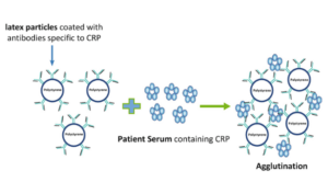
Polystyrene latex beads can be coated with antibody or antigen molecules that are randomly arranged. Since every latex particle has a massive amount of antibody or antigen molecules linked to it, there are many possible binding sites. To create cross-linked aggregates of latex beads and antigen or antibody, antigen or antibody in a specimen attaches themselves to combining sites on the latex beads that are exposed to the antigen/antibody. Due to the huge particle size of latex, it is possible to see all the antigen-antibody response in a clearer manner.
The Latex Agglutination Test Requirements
The Vials are 1.5 ml in size If you’re looking for microcentrifuges, pipettes and microtips, we’ve got you covered. Refrigeration in a laboratory Blocking buffer, Glycine saline buffer coating antigen Beads of latex a test antiserum Using a beaker, glass slides, and a toothpick.
Test for latex agglutination procedure
Latex microbeads are coated with pathogen-specific antigens or antibodies in both types of latex tests. This is done by serially diluting the patient’s cerebrospinal fluid, serum, or urine with the coated latex particles, and then watching for agglutination (clumping).
Latex Coatings (For detection of antibodies)
- You’ll need to add 40 μl of Glycine-Saline buffer to 20 μl latex beads.
- 2 hours at 37oC with 60μl of the antigen added to the latex.
- 5000 rpm for 10 minutes, then aspirate the supernatant with caution.
- You’ll need to disperse the pellet for 10 minutes in blocking buffer (1ml) before spinning it down.
- Laundering should be done a second time.
- To the pellet, add 90 l of blocking buffer, and mix thoroughly.
- Overnight, incubate at 4o C.
Agglutination Test
- Add 4 l of test antisera to 200 l of glycine-saline buffer in a vial. diluted by 50 times before using.
- Antigen is mixed with diluted serum in a 1.5-mL vial, and the mixture is incubated for 10 minutes at room temperature.
- Inject 10 l of a glass slide with latex.
- Slide A should have 10 l of diluted test antiserum.
- 10 l antiserum-antigen (from step 1) is added to B.
- Assemble C by adding 10l of glycine-saline buffer (G-S).
- To mix the content in each slide, use a toothpick. After using the toothpick in one slide, throw it away (take a new one for the next slide).
- When you’re done mixing, let it sit for 2 minutes to see what happens.
Results of Latex Agglutination Test and its Interpretation
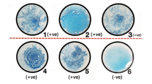
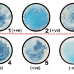
As a result of the method, certain reactions are recorded as positive or negative, while others are rated on a scale from 1 to 4, where two being the minimal quantity of agglutination observable even without a microscope.
A positive outcome is when the beads form clumps in any of the dilutions.
You can use it to determine whether or not a patient’s body has created a pathogen-specific antibody (if you used an antigen) or whether or not a sample contains the pathogen’s antigen (if the test supplied the antibody).
Negative: There is no clumping or agglutination.
Deficiency in the presence of pathogen-specific antigen or antibody.
Latex Agglutination Test in the Workplace
- In clinical laboratories, latex tests are often used to detect antigens to Cryptococcus neoformans in cerebral fluid or serum, respectively. When detecting capsular antigens of Pneumococcus, H. influenzae and Meningococcal bacteria it is also employed.
- Beta-hemolytic Streptococcus must be confirmed in culture plates.
- For example, Clostridium difficile toxin A and B are detected by latex testing, as is rotavirus and Escherichia coli 0157:H7 from questionable E coli colonies.
- C-reactive protein is most commonly detected by the latex agglutination test, which uses latex particles coated with anti-CRP antibodies. In the latex agglutination test, the detection limit for CRP is 0.6 mg/dl.
- Test for Antibody Detection Using Latex Agglutination (antistreptolysin O antibody).
Latex Agglutination Test Advantages
- 0.8 m or larger latex beads make it easier to see the agglutination response.
- The LAT is one of the most extensively utilised tests today since it is quick and simple to administer and interpret. One theory holds that it has supplanted many other serological tests, such as co-agglutination assays.
- As well as being reasonably priced and relatively stable, they do not cross-react with other antigens.
- Latex agglutination has been proven to identify bacterial polysaccharides as low as 0.1 ng/mL.
Latex Agglutination Limitations
- There must be strict standards in place for latex agglutination operations, because factors such as pH, osmolarity and ionic concentration have an impact on the quantity of binding that happens.
- As an example, the rheumatoid factor has been reported to induce false positive reactions in the latex agglutination systems.
Before testing, several agglutination procedures involve the specimens to be heated at 56°C or treated with EDTA, which is a time-consuming process
Latex Agglutination Citations
- Alberts, B. (2004). Essential cell biology. New York, NY: Garland Science Pub.
- Kar,D.K. and halder,S. (2015). Cell biology genetics and molecular biology.kolkata, New central book agency
- https://www.ncbi.nlm.nih.gov/books/NBK9886
- https://micro.magnet.fsu.edu/cells/microfilaments/microfilaments.html
- https://biologydictionary.net/microfilament/
- https://www.sciencedirect.com/science/article/pii/B9780080552323600510
Related Posts
- Anisocytosis: Definition, Types, Causes, Symptoms, Treatment
- Endospore Staining: Principle, Procedure, Reagents, Results
- Flow Cytometry: Overview, Principle, Steps, Types, Uses
- Northern Blot: Overview, Principle, Procedure and Results
- MPV Blood Test: Calculation, High and Low MPV Value, Results
- Latex Agglutination Test: Objectives, Principle, Procedure, Results
- Iodine Test: Definition, Objective, Principle, Procedure, Results
- Eosin Methylene Blue (EMB) Agar
- Biuret Test for Protein: Purpose, Objectives, Principle, Procedure, Reagents
- Streak Plate Method: Meaning, Principle, Methods, Importance, Limitations
- Bile Solubility Test: Objective, Principle, Procedure, Results, Uses
- Butyrate Disk Test: Objective, Principle, Procedure, Results, Uses, Limitations
- Beta Lactamase Test: Objective, Principle, Procedure, Results, Limitations
- Bacitracin Susceptibility Test: Objective, Principle, Procedure, Results, Uses, Limitations
- Bile Esculin Test: Objective, Principle, Procedure, Result, Uses, Limitations

