Definition of Microtubules (What are Microtubules?)
- Microtubules are located within the cytoplasm of every eukaryotic cells, with the exception of human erythrocytes.
- Microtubules are small, empty, bead-like tubular structures that aid in cell shape maintenance.
- These are small hollow tubes present within cells that also serve as the cell’s motor.
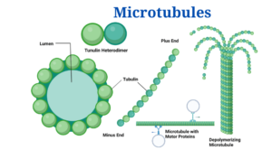
Figure: Diagram of Microtubules Image Created with BioRender
Microtubule Structure
- These are lengthy fibres (indefinite length) with a diameter of 24 nm.
- Every microtubule seems to contain a dense wall of 6 nm thickness plus a light or empty centre in cross-section. A microtubule’s wall is composed of 13 globular subunits termed protofilaments that are around 4 to 5 nm in diameter.
- Chemically, they are made up of two types of protein subunits: -tubulin (tubulin A) and -tubulin (tubulin B), every with a molecular weight of 55,000 daltons.
- A microtubule’s wall is formed of a helical array of repeated and tubulin subunits.
- According to assembly studies, the structural unit is an 8 nm long dimer.
- Therefore, every microtubule contains 13 protofilaments, each of that is made up of dimers that run parallel to the tubule’s lengthy axis. The repeating unit is a heterodimer that is oriented ‘head to tail’ inside the microtubule, i.e.
- As a result, every microtubule has a distinct polarity: its two ends are not structurally similar.
Assembly
- Microtubules go through reversible assembly and disassembly (i.e., polymerization and depolymerization) in response to the needs of the cell or organelles.
- Some microtubule-associated proteins control their polymerization (MAPs).
- Microtubule assembly includes the privileged adding up of subunits (dimers) to one ending of the tubule, known as the A end (or net assembly end); the opposite ending of the tubule is known as the D end (or net disassembly end). The hydrolysis of GTP to GDP is part of such an assembly. As a result, tubulin assembly in the creation of microtubules is a highly orientated and regulated process.
- This assembly is oriented at centrioles, basal bodies, and centromeres of chromosomes. Calcium and calmodulin (an acidic protein with four Ca2+ binding sites) are two other variables that influence tubulin polymerization in vivo.
Microtubules can function alone or in collaboration with other proteins to generate extra complex structures. Cell organelles generated from unique microtubule assemblies involve:
- Cilia and flagella are two types of flagella.
- Centrioles and basal bodies
Microtubules Functions
- They use unique attachment proteins to transport vesicles, granules, organelles such as mitochondria, and chromosomes.
- They, along with microfilaments and intermediate filaments, compose the cytoskeleton of the cell and play various motor tasks for the cell.
Microtubules, as part of the cell’s cytoskeleton, help with:
- Cells and cellular membranes are given shape.
- Cell movement, that involves muscle cell contraction as well as other processes.
- Microtubule “roadways” or “conveyor belts” transport certain organelles within the cell.
- Mitosis and meiosis are the processes by which chromosomes migrate during division of cell and the mitotic spindle is formed.
Microtubules Citations
- Verma, P. S., & Agrawal, V. K. (2006). Cell Biology, Genetics, Molecular Biology, Evolution & Ecology (1 ed.). S .Chand and company Ltd.
- Alberts, B. (2004). Essential cell biology. New York, NY: Garland Science Pub.
- Kar,D.K. and halder,S. (2015). Cell biology genetics and molecular biology.kolkata, New central book agency
- http://cytochemistry.net/cell-biology/microtub.htm
- https://sciencing.com/main-function-microtubules-cell-8552402.html
- http://www.softschools.com/science/biology/the_function_of_microtubules/
Related Posts
- Phylum Porifera: Classification, Characteristics, Examples
- Dissecting Microscope (Stereo Microscope) Definition, Principle, Uses, Parts
- Epithelial Tissue Vs Connective Tissue: Definition, 16+ Differences, Examples
- 29+ Differences Between Arteries and Veins
- 31+ Differences Between DNA and RNA (DNA vs RNA)
- Eukaryotic Cells: Definition, Parts, Structure, Examples
- Centrifugal Force: Definition, Principle, Formula, Examples
- Asexual Vs Sexual Reproduction: Overview, 18+ Differences, Examples
- Glandular Epithelium: Location, Structure, Functions, Examples
- 25+ Differences between Invertebrates and Vertebrates
- Lineweaver–Burk Plot
- Cilia and Flagella: Definition, Structure, Functions and Diagram
- P-value: Definition, Formula, Table and Calculation
- Nucleosome Model of Chromosome
- Northern Blot: Overview, Principle, Procedure and Results

