Monocot Leaves Definition
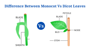
Monocotyledonous plants have narrow, elongated leaves with parallel venation, which is used to distinguish them from dicots.
- Monocot leaves are isobilateral because the colour on both surfaces of the leaves is same.
- A proximal leaf base, or hypophyll, and a distal leaf hyperphyll make up the primordial monocot leaves. In dicots, the hyperphyll is the major structure of the leaf, but in monocots, the hypophyll is the dominant structure.
- As previously stated, the venation is of the striate type, which is primarily longitudinally striate but can also be palmate-striate or pinnate-striate.
- The veins on the leaf surface start at the base and travel up to the apex.
- Because the leaf base comprises more than half of the circumference of the plant stem, most monocotyledonous plants have just one leaf per node.
- Differences in stem development during zonal differentiation have been blamed for the existence of a bigger leaf base.
Dicot Leaves Definition
Dicotyledonous leaves are usually rounded, with reticulate venation, and are structurally and anatomically distinct from monocotyledonous leaves.A lead blade, also known as the lamina, is found on a typical dicot leaf.The lamina is the leaf’s broadest section.
- Dicot leaves are dorsoventral because the coloration of the dorsal and ventral parts of the leaves may be distinguished. The leaf’s dorsal side is frequently more coloured than its ventral side.
- In contrast to monocot leaves, which are directly linked to the stem, dicot leaves contain a petiole that connects them to the stem.
- Some dicot leaves have little green appendages called stipules at the base of the petiole.
- The midrib of dicot leaves goes through the leaf blade and runs the length of the leaf.
- The reticulate venation is formed when several branches emerge on either side of the midrib. The number of leaves on a stem node varies per species, but dicots typically have two or more leaves emerging from a single node.
- Dicot leaves are further classified into categories based on the leaf morphology, with some having simple leaves and others having compound leaves.
Leaves of Monocots and Dicots have different structures.
The following structures can be used to describe the internal structure or anatomy of monocot and dicot leaves:
- The epidermis.
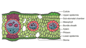
- The epidermis is a dense layer of thin-walled barrel-shaped cells that makes up the outermost tissue in leaves. On both the upper and lower parts of the leaf, the epidermis is present. The epidermis is protected by the cuticle, which is a waxy covering that protects the leaf and prevents water loss.
- The cuticle on the upper layer of the leaf is thicker than that on the lower surface in dicot leaves as the leaves are dorso-ventral.Monocots, on the other hand, have an epidermal layer that is almost the same thickness on both surfaces.
- The epidermis is necessary because it is a waxy waterproof layer that inhibits the loss of surplus water. Furthermore, because it contains microscopic pores known as stomata, it plays a crucial function in gas exchange.
- In monocot leaves, the number of stomata is identical on both surfaces. In dicot leaves, the lower epidermis has more stomata than the top epidermis.
- Stomata are small openings that regulate the size of the stomata. They are found between bean-shaped cells called guard cells. In dicots, the guard cells are dumb-bell shaped.
- The epidermis also contains subsidiary cells, which are typically found around the guard cells.
- In the case of dicot cells, the upper epidermis contains large thin-walled cells called bulliform cells, which contain chloroplasts that give the leaves their characteristic green colour. These cells help the leaves roll in response to changes in the weather.
- In monocot leaves, some epidermal cells are filled with silica, which are known as silica cells.
- Mesophyll
- The mesophyll is the leaf’s ground tissue that lies between the upper and lower epidermis.
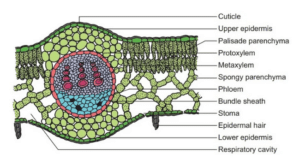
- In dicot leaves, the mesophyll is divided into palisade and spongy parenchyma, whereas in monocot leaves, no such separation exists.
- The palisade parenchyma is made up of vertically elongated cylindrical cells in one or more layers and is found directly under the top epidermis. There are no intercellular gaps because the cells are packed tightly together.
- The palisade parenchyma’s cells have more chloroplasts than the spongy parenchyma’s cells.
- The spongy parenchyma is found beneath the palisade parenchyma, with irregularly structured cells.There are fewer chloroplasts in these cells than in palisade parenchymal cells.
- The spongy parenchyma’s cells are loosely packed with many gaps, hence the name.
- The exchange of gases between the cells is facilitated by these air gaps. Respiratory cavities, also known as sub-stomatal cavities, are the spaces between the stomata.
- The dicot leaves’ mesophyll is made up of isodiametric and thin-walled cells that are not divided into palisade and spongy parenchyma.
- With some intercellular air gaps, the cells are compactly organised.
Figure: T.S. of a dicot leaf (sun-flower). Image Source: BrainKart.
- Vascular Bundles
- The vascular bundle, which runs beneath the mesophyll and along the veins of the leaves, is the innermost layer of tissues in plant leaves.
- The vascular bundles are not all the same size, and the larger bundles are spaced out along the veins at regular intervals. Above and below the major vascular bundles, there are two patches of sclerenchyma cells.
- The vascular bundles of the leaves are part of the plant’s vascular system, which runs through several organs and encompasses the entire plant.
- In terms of size and position, the vascular bundles in leaves have very different patterns.
- The veins of dicot leaves are of varying sizes, generating a highly branching network. As a result, the vascular bundles found beneath such veins are different.
- Monocot leaves have longitudinal veins that run parallel to the leaf blade and are joined transversally by tiny commissural veins, resulting in vascular bundles that are less varied.
- A bundle sheath surrounds the vascular bundles, which are conjoint, collateral, and closed.
- Dicot leaves’ bundle sheaths are parenchymatous, but dicot leaves’ sheaths are sclerenchymatous.
- The vascular bundle’s xylem tissue is found in the upper epidermis, while the phloem tissue is found in the lower epidermis.
- Vessels and xylem parenchyma make up the xylem.In monocot leaves, the xylem is divided into metaxylem and protoxylem. Sieve cells, companion cells, and phloem parenchyma make up the phloem.
Functions of Monocot and Dicot Leaves
The functions of leaves are essentially identical in both dicot and monocot plants. The functions of leaves are influenced by the plant’s species, environment, and age. Some of the roles of monocot and dicot plants are as follows:
- The preparation of food via the process of photosynthesis is the most significant function of leaves in green plants. As one-fifth of the cells in the mesophyll contain chlorophyll-containing chloroplasts, the cells in the leaves contain chlorophyll. The leaves’ big, broad surface allows more sunlight to be absorbed, which is necessary for photosynthesis.
- The epidermis and cuticle of the leaves keep the plant from drying out by preventing excessive loss during transpiration.
- Stomata, which are a necessary structure for the passage of water from the plant into the atmosphere via the transpiration process, are found on the leaves. This is necessary in order for the roots to take water containing minerals from the soil.
- Stomata are engaged in the exchange of gases as well as promoting transpiration. During photosynthesis, the stomata take in carbon dioxide from the air and release oxygen.
- Food created by the cells in the leaves of some plants, such as cabbage and lettuce, is stored in various ways in the leaves.
Difference Between Monocot and Dicot Leaves
Monocot vs Dicot Leaves
| Characteristics | Monocot leaves | Dicot leaves |
| Definition |
Monocotyledonous plants have narrow, elongated leaves with parallel venation, which is used to distinguish them from dicots. |
Dicotyledonous leaves are usually rounded, with reticulate venation, and are structurally and anatomically distinct from monocotyledonous leaves. |
| Shape |
The leaves of monocots are narrower, slenderer, and longer than those of dicots. |
The leaves of dicots are wider and smaller than those of monocots. |
| Symmetry |
The symmetry of monocot leaves is isobilateral. |
Because the upper and lower surfaces of the leaves are distinct, dicot leaves are dorsoventral. |
| Venation |
The longitudinal veins traverse the length of the leaf and are joined by microscopic commissural veins in monocot leaves, which have parallel venations. |
The reticulate venation of dicot leaves is made up of veins of various sizes that are joined to form a complicated network. |
| Stomata |
Monocot leaves are also known as amphistomatous because the number of stomata on the upper and lower surfaces of the leaves is equal. |
The lower surface of dicot leaves has more stomata than the top surface. Some dicot leaves lack stomata on the upper surface, and these plants are known as hypostomatous. |
| Guard cells |
The guard cells of monocot leaves are formed like dumbbells. |
Kidney-shaped guard cells can be seen in dicot leaves. |
| Intercellular spaces |
Monocot leaves contain reduced intercellular gaps due to the tight arrangement of the cells. |
Because the cells in dicot leaves are loosely packed, there are more intercellular gaps. |
| Vascular bundles |
The monocot leaves have both big and small vascular bundles. |
The vascular bundles of dicot leaves are bigger. |
|
Monocot leaves have two types of xylem: metaxylem and protoxylem. |
In dicot leaves, the xylem is not divided into metaxylem and protoxylem. |
|
|
The monocot leaves’ bundle sheath is sclerenchymatous. |
The dicot leaves’ bundle sheath is parenchymatous. |
|
| Epidermis |
Silica is heavily deposited in the epidermal cells of monocot leaf epidermal cells. |
There is no silica deposition in the epidermal cells of dicot leaves. |
|
Monocot leaves have bulliform or motor cells in their epidermis. |
Bulliform and motor cells are absent from the epidermis of dicot leaves. | |
| Mesophyll |
Monocot leaves have two types of mesophyll: spongy mesophyll and palisade mesophyll. |
Dicot leaves have no distinct mesophyll. |
Click Here for Complete Biology Notes
Monocot Leaves Examples:
- Maize leaves
- Maize leaves are the most common monocot leaves, with basic, ordered structures.
- A normal maize plant has roughly 20 leaves, which might be in various states of development. A ripe maize leaf measures 70 cm in length and 8 cm in width.
- The top blade, lower sheath, and auricle are the three parts of a maize leaf. Leaf cells are orientated and arranged in such a way that they produce parallel lines and an elongated shape.
- The mesophyll and vascular tissue are encased by adaxial and abaxial epidermal tissue in the maize leaf blade.
- There are two types of cells in the maize leaf epidermis: specialised cells and nonspecializedintercostals cells.
- Stomatal complexes and three types of hair cells, enormous macrohairs, microbars, and bicellular hairs, make up specialised cells. These cells have a protective role to play.
- Grass
A grass leaf is a monocot leaf with an extended structure that develops from the node and is made up of a basal cylindrical sheath that encircles the stem and other younger leaves.
- On the outside, the sheaths are hollow cylinders that divide down one side. Typically, these sheaths form overlapping formations.
- The leaf has an auricle, which can be an ear-like projection or a hairy edge near the base of the leaf blade.
- The leaf has an auricle, which can be an ear-like projection or a hairy edge near the base of the leaf blade.
- A grass leaf’s epidermis is made up of specialised hair cells that defend the plant from different hazardous substances.
- Because the shape, texture, and hairiness of grass leaves vary between species and even within the same plant, they are frequently utilised as diagnostic criteria for grass distinction.
Examples of Dicot Leaves
- Mustard leaves
- Mustard leaves are an example of a dicot leaf.
- Mustard is a common dicot plant that is utilised in dicotyledonous plant research.
- Mustard leaves begin to grow in 4 weeks, when they reach a length of 6-8 inches. In around 6 weeks, a mature mustard leaf will be 15-18 inches long.
- Green leaves with reticulate venation are broad and green. The leaves are flattened dorsoventrally, with the ventral surface being lighter than the dorsal.
- Mustard leaves have a few unique hair cells in their epidermis that protect them against water loss and toxic substances.
- Mustard leaves are utilised as vegetables because they are high in nutrients that are beneficial to human health.
- Mint leaves
- Mint is a rapidly growing plant with round lanceolate leaflets placed in opposing pairs on the stem.
- The leaves are tiny, ranging in length from 4 to 5 inches. The size of the leaves, on the other hand, is determined by their age and maturity.
- Reticulate venation is seen on the leaves, with a central midrib from which branched veins emerge.
- The leaves’ upper and lower surfaces are coated in microscopic hairs.
- Mint leaves are fragrant and have a distinct mint scent. These leaves are utilised in a variety of foods in various civilizations.
Monocot Leaves Vs Dicot Leaves Citations
- Sylvester A.W., Smith L.G. (2009) Cell Biology of Maize Leaf Development. In: Bennetzen J.L., Hake S.C. (eds) Handbook of Maize: Its Biology. Springer, New York, NY. https://doi.org/10.1007/978-0-387-79418-1_10
- https://courses.lumenlearning.com/boundless-biology/chapter/leaves/
- https://vivadifferences.com/monocot-leaf-vs-dicot-leaf/
- https://nph.onlinelibrary.wiley.com/doi/full/10.1111/j.1469-8137.2004.01191.x
- https://en.mimi.hu/biology/lower_epidermis.html
- https://de.answers.yahoo.com/question/index?qid=20060909022759AAG460M
- https://botit.botany.wisc.edu/botany_130/anatomy/Leaf/Leaf.html
- http://mahatmaacademy.com/uncategorized/ncert-botany-anatomy-flowering-plant-online-model-paper/
- https://www.visiblebody.com/learn/biology/monocot-dicot/leaves
- https://www.vedantu.com/biology/difference-between-monocot-and-dicot-leaf
- https://www.thoughtco.com/plant-stomata-function-4126012
- https://www.sciencedirect.com/science/article/pii/S167420521500204X
- https://www.sciencedirect.com/science/article/pii/0020740381900539
- https://www.quora.com/What-is-the-arrangement-of-leaves-on-a-stem
- https://www.medicinenet.com/epidermis/definition.htm
- https://www.differencebetween.com/difference-between-upper-and-vs-lower-epidermis/
- https://www.diffen.com/difference/Dicot_vs_Monocot
- https://www.britannica.com/science/mesophyll
- https://www.bioexplorer.net/epidermis-cells.html/
- https://www.answers.com/Q/Why_do_palisade_mesophyll_cell_contains_more_chloroplast_than_the_spongy_mesophyll_cell
- https://www.answers.com/Q/What_is_the_function_of_the_leaf_sheath_in_the_monocotyledon_plant
- https://wikimili.com/en/Monocotyledon
- https://vivadifferences.com/difference-between-protoxylem-and-metaxylem/
- https://study.com/academy/lesson/structure-of-leaves-the-epidermis-palisade-and-spongy-layers.html
Related Posts
- Phylum Porifera: Classification, Characteristics, Examples
- Dissecting Microscope (Stereo Microscope) Definition, Principle, Uses, Parts
- Epithelial Tissue Vs Connective Tissue: Definition, 16+ Differences, Examples
- 29+ Differences Between Arteries and Veins
- 31+ Differences Between DNA and RNA (DNA vs RNA)
- Eukaryotic Cells: Definition, Parts, Structure, Examples
- Centrifugal Force: Definition, Principle, Formula, Examples
- Asexual Vs Sexual Reproduction: Overview, 18+ Differences, Examples
- Glandular Epithelium: Location, Structure, Functions, Examples
- 25+ Differences between Invertebrates and Vertebrates
- Lineweaver–Burk Plot
- Cilia and Flagella: Definition, Structure, Functions and Diagram
- P-value: Definition, Formula, Table and Calculation
- Nucleosome Model of Chromosome
- Northern Blot: Overview, Principle, Procedure and Results

