What is Microscope: Microscope Overview
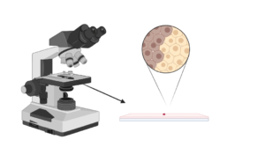
- As the name implies, microanalysis is the study of instruments (microscopes) used to view things or things that can’t be seen with open eyes.
- Microscopes use physical magnification to magnify the image of an object to make it visible.
- Microscopic substances are those that can only be seen under a microscope.
- Studies in microbiology, which deals with the structure and function of microscopic organisms, require magnifying glasses and microscopes.
- Subcellular organelles such as the nucleus and chromosomes can be seen using X-ray microscopy, a relatively recent imaging method.
- When Anton Von Leeuwenhoek invented the first microscope, it marked the beginning of modern microscopy, particularly optical microscopy.
- With the advent of new and improved microscopes with higher magnification and resolution, microscopy has become increasingly difficult since then.
- The optical microscopes magnify objects by passing light through a series of glass lenses into the observer’s eyes to create a magnified image.The most popular type of microscope is a ‘compound microscope,’ which is mainly used for research and education.
- An electron and X-ray microscope uses an electron beam to create a digitally enlarged image of whatever it looks at.
- With ‘electron microscopes,’ you can see objects as small as an atom enlarged thanks to their great magnification and resolution.
- Different microscopes, such as bright field, fluorescence, phase contrast, and darkfield microscopes, are accessible depending on the sample’s nature
- .As a result, the magnification and resolution of the pictures produced by a given microscope are dependent on the lens type utilized in the system.
- Histology, cytology, and bacteriology all rely heavily on microscopy.
- An essential technique for identifying microorganisms has long been a microscopic examination of cells’ morphology and structural characteristics.
What a microscope does?
When employed in science laboratories, microscopes provide a contrasted view of microscopic objects, such as cells and bacteria, by being magnified.
What is the purpose of Microscope?
It is commonly used to examine minute organisms like algae and fungi, and biological specimens under the microscope.
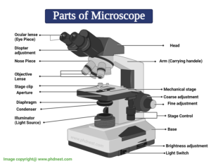
Diagram of Microscope
Figure: Parts of a Microscope
Parts of a Microscope and their Functions
The microscope comprises three main structural components: the head, the base, and the arm.
HEAD: Also known as the body, the microscope’s head houses the optical components in the upper portion.
BASE- It serves as a support for microscopes. Illuminators for microscopic work are also included. In the microscope, the amount connects the eyepiece tube and the base:
ARMS. Besides supporting the microscope’s head, it also serves as a handle for transporting the instrument. For better viewing, some high-end microscopes have an articulated arm with multiple joints. This allows the microscope head to move more freely.
Optical Components of Microscope
To see, magnify, and create a picture of a specimen on a slide, a microscope’s optical components must be utilized. Included in this list are the following elements:
1 . Eyepiece : It is also known as the ocular, is the portion of the microscope through which you view the specimen under examination.
2. Tube for the eyepiece: A holder for an eyepiece is precisely what it sounds like. The eyepiece is located above the objective lens.
3. For specimen visualization, the most common lenses employed are objective lenses.
The rotating turret is another name for the nose piece. It houses the telescope’s lenses.
4. The knobs : It is for adjusting the microscope’s focus are located here.
5. Specimen viewing section: this is where the specimens will be displayed for the audience to see.
6. The rotating turret: It is another name for the nose piece. It houses the telescope’s primary optics. Because it is moveable, the objective lenses can be swivelled to adjust for magnification power.
7. Aperture: It is the microscope stage’s hole through which light is transferred.
8. Illuminator: The light source for a microscope is called an illuminator.In illuminators, condensers are lenses that gather and focus light.
9. Iris: It is also called the diaphragm and has the primary function of controlling how much light gets to the specimen.
10. Illuminator Condenser Lenses : They are used to collect and focus light onto the specimen. Microscope diaphragms and stages are located under the stage. In order to obtain clean and clear images at magnifications of 400X and higher, they are crucial. Clearer images are obtained by using a magnifier with a greater magnification setting on the camera.
11. Diaphragm: The Abbe condenser, which is found in more advanced microscopes, magnifies objects by around 1000 times their original size.The diaphragm is another name for the iris, which is its more common scientific name.Located behind the microscope stage, its principal function is to regulate the amount of light that reaches the specimen under investigation. Being an adjustable device, the intensity and size of the light beam emitted to the specimen can be customised. These two components, which are found on high-quality microscope diaphragms, allow the microscope operator to fine-tune light intensity while also controlling the microscope’s overall focus.
12. Condenser focus knob: Controls the focus of light on the specimen by moving the condenser up or down in relation to the control knob for condenser elevation.
13. The Abbe Condenser : It is a condenser built specifically for high-quality microscopes, which allows the condenser to be moved and has very high magnifications of over 400X. In general, the numerical aperture of high-quality microscopes is greater than that of the objective lens.
14. The rack stop: In order to prevent the objective lens from going too close to the specimen slide and damaging it, it has a rack stop. It protects the objective lens from being damaged by the specimen slide rising too high.
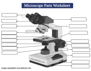
Microscope Labeled Diagram
Revision Questions : FAQ
How to tell the difference between a standard condenser and an Abbe condenser?
Using a condenser, the illuminator’s light can be collected and focused upon a specimen. Under the microscope stage, near the diaphragm, they can be found. They are critical in obtaining crisp, clear images at magnifications of 400X and higher. If you have a high-quality microscope that comes with an Abbe condenser, the condenser can be moved around the microscope, allowing you to magnify objects at magnifications up to 400X. In general, the numerical aperture of a high-quality microscope is greater than that of an objective lens.
When comparing the eyepiece and objective lens, how large is the objective lens?
The part of the microscope through which you view things is called an ocular or an eyepiece. It’s located at the very top of the microscope, as the name implies. An extra eyepiece with a magnification range of 5X – 30X can be added to the scope’s regular 10x magnifier. The most common type of lens used for specimen visualisation is an objective lens. They can magnify objects 40x to 100x their original size. Depending on the microscope, there can be anywhere from one to four objective lenses. Some are backward-facing while others are forward-facing.
It’s possible to change the rack stop, so why do microscopes come with it?
In order to avoid damaging the objective lens, a rack stop is installed in the microscope.
Microscopes Citations
- https://www.pobschools.org/cms/lib/NY01001456/Centricity/Domain/349/TheMicroscope-howtouse.pdf
- https://sciencing.com/parts-microscope-uses-7431114.html
- https://www.amscope.com/microscope-parts-and-functions/
- https://cpb-us-e1.wpmucdn.com/cobblearning.net/dist/3/4204/files/2018/08/Parts-of-the-Microscope-103b21p.pdf
Related Posts
- Phylum Porifera: Classification, Characteristics, Examples
- Dissecting Microscope (Stereo Microscope) Definition, Principle, Uses, Parts
- Epithelial Tissue Vs Connective Tissue: Definition, 16+ Differences, Examples
- 29+ Differences Between Arteries and Veins
- 31+ Differences Between DNA and RNA (DNA vs RNA)
- Eukaryotic Cells: Definition, Parts, Structure, Examples
- Centrifugal Force: Definition, Principle, Formula, Examples
- Asexual Vs Sexual Reproduction: Overview, 18+ Differences, Examples
- Glandular Epithelium: Location, Structure, Functions, Examples
- 25+ Differences between Invertebrates and Vertebrates
- Lineweaver–Burk Plot
- Cilia and Flagella: Definition, Structure, Functions and Diagram
- P-value: Definition, Formula, Table and Calculation
- Nucleosome Model of Chromosome
- Northern Blot: Overview, Principle, Procedure and Results

