Definition of Voluntary Muscles
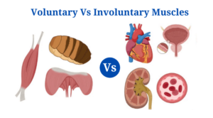
Voluntary muscles are those that can be moved by a person’s free will and are virtually always linked to the skeletal system.
- These muscles are responsible for all types of motions in vertebrates and are connected to the bones via tendons.
- Voluntary muscles make up around 40% of the body’s total weight and are usually lengthy and located near the bones.
- Because voluntary muscles are made up of long, thin, and multinucleated muscle fibres that are crossed with a regular pattern of red and white red and white lines, they have a striated appearance.
- Each muscle cell is nucleated, with the nucleus remaining on the cell’s perimeter.
- A unique cell membrane called the myolemma or sarcolemma surrounds the muscle fibres.
- The sarcolemma is a thick connective tissue that joins muscle fibres and connective tissues in voluntary muscles.
- Furthermore, sarcomeres, which are contractile units in muscle fibres, shorten, causing the muscle to contract and relax.
- Actin and myosin proteins are found in sarcomeres, and they work together to cause muscle contraction by sliding against each other.
- Connective tissue connects muscle fibres, which communicate with one another via nerves and blood arteries.
- Afferent and efferent nerves make up the somatic nervous system, with afferent nerves relaying information to the central nervous system and efferent nerves relaying information from the CNS to voluntary muscles for contraction.
- These muscles are not myogenic, and they require a nerve signal from outside to contract.
- The contraction and relaxation of voluntary muscles demand a significant amount of energy. As a result, they have many mitochondria to suit their energy requirements.
- In comparison to involuntary muscles, voluntary muscles contract and relax quickly. They do, however, get tired rapidly and need to rest at regular intervals.
- These muscles are crucial since they are involved in the movement of bodily components as well as the body’s mobility.
- The biceps, triceps, quadriceps, diaphragm, pectoral muscles, abdominals, hamstrings, and other voluntary muscles are examples.
Definition of involuntary muscles
Involuntary muscles are those that cannot be controlled by volition or conscious thought, and are frequently connected with organs that contract and relax slowly and regularly.
- Because there are no striations when studied under a microscope, involuntary muscles are also known as smooth muscles or non-striated muscles.
- Internal organs such as the stomach, colon, urine bladder, and blood capillaries have these muscles lining their walls.
- Individual smooth muscle cells are long, thin, and spindle-shaped, with a nucleus in the centre.
- The myolemma, also known as the sarcolemma, is the cell membrane that connects muscle fibres together. The sarcolemma present is thinner and has a lower concentration.
- The cardiac muscle is an example of an involuntary muscle that differs in structure and function from other involuntary muscles.
- Individual heart muscle cells termed cardiomyocytes are connected together by intercalated discs in the cardiac muscle. Collagen fibres and other components that make up the extracellular matrix surround these muscle cells.
- The contraction of cardiac muscle differs from skeletal and smooth muscle contractions. Electrical stimulation causes the action potential to be formed within the muscles.
- Calcium ions are released from the cells into the sarcoplasm reticulum as a result of this potential. The myofilaments move past each other as calcium ions rise, generating excitation-contraction.
- The nerve stimulus is created within the muscles of the cardiac muscle, making it myogenic.
- The majority of muscle cells within the involuntary muscles’ muscle fibres work as a single unit, contracting and relaxing simultaneously.
- The autonomous nervous system of the peripheral nervous system controls involuntary muscles.
- Varicosities are neurotransmitter-filled bulges found in the motor nerves of the autonomous nervous system.
- Neuron signals can be transmitted from one cell to another via neurotransmitters because gap junctions connect the cells in involuntary muscles.
- Involuntary muscular contractions and relaxations are slow and occur at regular intervals.
- As a result, these muscles do not become fatigued easily and can work indefinitely.
- They also demand less energy than voluntary muscles, which means they have fewer mitochondria.
- Internal organ motions are aided by involuntary muscles, which also aid in the transport of fluids and food through the digestive system.
- The heart muscle and smooth muscle lining the intestinal tracts, blood arteries, urogenital tracts, respiratory tract, and other areas are examples of involuntary muscles.
Click Here for Complete Biology Notes
Important Differences Between Voluntary and Involuntary Muscles
[ninja_tables id=”3859″]Voluntary Muscles Examples
Diaphragm
- The diaphragm is a main respiratory muscle that helps with breathing by expanding and lowering the thoracic wall’s volume.
- It’s a dome-shaped skeletal muscle that lies beneath the lungs and heart. It separates the abdomen and chest regions.
- The phrenic nerve, which extends from the neck to the diaphragm, controls the diaphragm, which is a voluntary muscle.
- The esophageal aperture, which houses the vagus nerve, the aortic opening, which houses the aorta, and the caval opening, which houses the inferior vena cava, are all major openings in the diaphragm.
- The diaphragm is involved in a variety of non-respiratory processes in addition to respiratory ones. To aid in the elimination of vomit, pee, and faeces, the diaphragm raises abdominal pressure. It also creates esophageal pressure to avoid acid reflux.
- The sound of hiccupping is produced by spasmodic inspiratory movement of the diaphragm.
Biceps
- Bicep muscles have two heads or places of origin in humans, which are the bicep brachii and bicep femoris.
- The bicep brachii muscle is located on the front of the upper arm. Its tendons attach to the inner projection near the head of the radius, which is one of the forearm’s two bones.
- The biceps brachii is a muscle that bends the forearm toward the upper arm and is utilised for lifting and tugging.
- The bicep brachii is said to be a symbol of bodily strength because of its size.
- One of the muscles on the back of the thighs is the bicep femoris.
- It is linked to the head of the fibula and tibia and arises from the back of the isthmus and the back of the femur.
- It is engaged in thigh movement as well as knee flexion.
Involuntary Muscles Examples
Cardiac muscle
- The cardiac muscle is an involuntary striated muscle that lines the inside of the heart and contracts and relaxes at regular intervals.
- Individual heart muscle cells termed cardiomyocytes are connected together by intercalated discs in the cardiac muscle. Collagen fibres and other components that make up the extracellular matrix surround these muscle cells.
- The contraction of cardiac muscle differs from skeletal and smooth muscle contractions.
- The nerve stimulus is created within the muscles of the cardiac muscle, making it myogenic.
- Electrical stimulation causes the action potential to be formed within the muscles.
- Calcium ions are released from the cells into the sarcoplasm reticulum as a result of this potential. The myofilaments move past each other as calcium ions rise, generating excitation-contraction.
- The contractions of the muscle fibres of cardiac muscles are controlled by vagal and sympathetic nerves.
Smooth muscle
- Smooth muscle is a nonstriated, involuntary muscle that is made up of single-unit or unitary muscles as well as multiunit muscles.
- Smooth muscle lines the inside walls of internal organs such as the intestines, urinary tract, and blood arteries.
- The ciliary muscle is a smooth muscle in the eye that dilates and controls the iris, which changes the shape of the lens.
- Single-unit smooth muscles are those in which the complete muscle contracts and relaxes at the same time. As independent units, the multiunit muscles can contract and relax.
- Nerve fibres encircle muscle fibres and convey neurotransmitters in the form of vesicles termed varicosities or boutons.
Voluntary and Involuntary Muscles Citations
- https://www.encyclopedia.com/medicine/anatomy-and-physiology/anatomy-and-physiology/muscles
- https://www.differencebetween.com/difference-between-voluntary-and-vs-involuntary-muscles/
- https://www.britannica.com/science/ciliaris-muscle
- https://www.answers.com/Q/What_causes_the_release_of_calcium_ions_from_the_sarcoplasmic_reticulum
- https://www.answers.com/Q/Is_skeletal_muscle_controlled_by_the_Autonomic_Nervous_System
- https://teachmephysiology.com/nervous-system/synapses/action-potential/
- https://teachmeanatomy.info/thorax/muscles/diaphragm/
- https://quizlet.com/187770489/anatomy-and-physiology-final-review-multiple-choice-flash-cards/
- https://pediaa.com/difference-between-cardiac-skeletal-and-smooth-muscle/
- https://brainly.in/question/765738
- https://brainly.com/question/9057101
- https://bodytomy.com/voluntary-muscles
- https://bodytomy.com/organs-of-digestive-system
Related Posts
- Phylum Porifera: Classification, Characteristics, Examples
- Dissecting Microscope (Stereo Microscope) Definition, Principle, Uses, Parts
- Epithelial Tissue Vs Connective Tissue: Definition, 16+ Differences, Examples
- 29+ Differences Between Arteries and Veins
- 31+ Differences Between DNA and RNA (DNA vs RNA)
- Eukaryotic Cells: Definition, Parts, Structure, Examples
- Centrifugal Force: Definition, Principle, Formula, Examples
- Asexual Vs Sexual Reproduction: Overview, 18+ Differences, Examples
- Glandular Epithelium: Location, Structure, Functions, Examples
- 25+ Differences between Invertebrates and Vertebrates
- Lineweaver–Burk Plot
- Cilia and Flagella: Definition, Structure, Functions and Diagram
- P-value: Definition, Formula, Table and Calculation
- Nucleosome Model of Chromosome
- Northern Blot: Overview, Principle, Procedure and Results

